Browse THP Patient Cases

Hendren Wilms Tumor Patient #3

This 3-year-old boy was hospitalized December 1978 with a mass palpable in both right and left flanks. Intravenous pyelogram (IVP) was consistent with bilateral Wilms’ tumors. Chest films were normal.

Angiography showed both kidneys displaced by a mass but with relatively intact renal parenchyma. Decision was made to treat patient with preoperative chemotherapy with hope to save most of each kidney if possible during surgery later.

As seen in the following six figures, there was progressive shrinkage of the two tumors. 

Fig. 1. Intravenous Pyelogram (Select Image for High-quality Version). IVP December 1978 consistent with bilateral Wilms’ tumors. Lungs clear on chest film.

 Fig. 2. Aortagram (Select Image for High-quality Version). Aortography on hospital day 3. Both main renal arteries are normal although displaced.

Fig. 3. Angiograph (Select Image for High-quality Version). Left kidney pushed upward, and lying transversely, by the tumor below it. Note intact parenchyma. 

Fig. 4. Intravenous Pyelogram (Select Image for High-quality Version). IVP after 1 month of chemotherapy with Actinomycin D and Vincristine. Masses smaller.

Fig. 5. Intravenous Pyelogram (Select Image for High-quality Version). IVP after 2 months of chemotherapy. Almost normal calyceal architecture on the left side. Mass behind left kidney 2 inches (5.08 cm) in diameter. Mass at upper pole of the right kidney 5 inches (12.7 cm) in diameter. 

Fig. 6. Angiograph (Select Image for High-quality Version). Selective renal angiogram, left side before surgery.  About 2/3's of parenchyma judged to be normal. 

Surgical excision was performed in March 1979, 2.5 months after admission. Under anesthesia during preliminary cystoscopy ureteral catheters were passed up to each kidney to allow irrigation of saline up to each after removal of the tumor to check for calyceal leaks. The ureteral catheters were taped to an inlying Foley catheter to keep them in place.

A long transverse upper abdominal “frown” incision was made from one flank to the other in supine position. There was no tumor visible or palpable except in the flanks. The left side was approached first, reflecting the left colon downward. Gerota’s capsule was opened, staying free from the tumor, then about the size of a squash ball. It lay posteriorly at the renal hilum. The anterior aspect of the kidney and both upper and lower poles were free. All vessels were isolated. The kidney was packed in ice to cool it. The vessels were then cross clamped and it was cooled further to lengthen the ischemic time which would be safe. The tumor was then dissected from the normal kidney with a 5 mm. margin of normal renal parenchyma with the specimen. Scissors were used for this dissection, guiding the plane of dissection with the palpating left hand fingers. All transected renal vascular branches were clamped and over sewn.

Fig. 7. Left Kidney. The posterior side of the left kidney with the tumor remarkably smaller than before chemotherapy

Fig. 8. Defect in Left Kidney. After removing the tumor by scissor dissection. Clamp time for the renal vessels had been 17 minutes.

An assistant irrigated the left ureteral catheter to demonstrate two opening to be closed in the renal collecting system. An omental patch was taken from the greater curvature of the stomach to suture over the renal defect. The kidney was then put back into Gerota’s capsule. The flank was drained.

The right side was then exposed, reflecting the hepatic flexure of colon medially. The tumor was much larger, 5 inches (12.7 cm) in diameter and occupying the upper 2/3 rds of the kidney. An identical procedure was performed, after renal cooling. Curiously the cross clamp time of ischemia perchance was 17 minutes, the same as the left had been. Omentum was not as generous on the right. A flap of Gerota’s fascia and adjacent fat was used instead, as a pedicle flap to cover the renal open surface.

Fig. 9. Right Kidney. Kidney mobilized and draped, to cool and clamp. Tumor occupied the entire upper kidney.

Fig. 10. Tumor Removed. Renal pelvis to close in center of kidney. A pedicle flap of Gerota’s covered the defect.

Nitroprusside hypotension to 65 mm. Hg. Intraoperatively helped reduce blood loss during this 6-hour operation.

Fig. 11. Retrograde Injection of Ureteral Catheters. One week post-operatively demonstrating no leaks from right hemi kidney or nearly normal left kidney. 

Fig. 12. Intravenous Pyelogram. IVP two weeks post-operatively demonstrating good function on both sides.

In January 1980, at age 4 years the patient had an episode of intestinal obstruction from an adhesion across the terminal ileum.

Chemotherapy was continued for a total of two years. Radiation was never given because there was no suspicion of residual tumors.

The pathology examination described nephroblastoma with focal rhabdomyomatous differentiation.

At age 11 years he was in a motor scooter accident and had head trauma, requiring 3 months of hospitalization.

At age 13 years he was evaluated for endocrinopathy after excessive weight gain. He complained also of panic attacks. No endocrine problem was identified.

Hendren Wilms Tumor Patient #4

This 27-month-old boy was referred in December 1969 with a very large left-sided Wilms’ tumor. An older sister at age 3½ years had been operated on elsewhere with a large right-sided Wilms’ tumor in 1966. Despite radiation and chemotherapy, she died in 1967, one month after the birth of her younger baby brother.

Intravenous pyelogram of this boy showed nonvisualization of both renal collecting systems. An inferior vena cava study, with a greater volume of contrast, showed obstruction of the cava and extension of the left-sided tumor up the vena cava into the right atrium. Satisfactory visualization of the renal collecting systems on both sides was seen with the increased contrast bolus. Three pulmonary metastases were seen in the right lower lung field. Venous return from both kidneys was via collateral veins rather than the renal veins because of the caval obstruction by tumor. Liver chemistries suggested obstruction of hepatic venous vessels, also producing the Budd-Chiari Syndrome.

Fig. 1. Preoperative Chest Film Showing Diaphragm Pushed Upward by Ascites and Fluid in Right Thorax (Select Image for High-quality Version). Liver palpably enlarged. These findings were secondary to blockage of hepatic venous outflow by tumor in the inferior vena cava. (Budd-Chiari Syndrome)

Fig. 2. Vena Cava Angiograms (Select Image for High-quality Version). (A) Preoperative study showing complete obstruction of intrahepatic I.V.C. by tumor extending from left renal vein up into right atrium. Note extensive para spinal collateral veins. (B) Postoperative study showing open I.V.C. and free flow to right heart after removal of left Wilms’ tumor and extraction of tumor previously blocking I.V.C. Normal I.V.P. right kidney. Liver enlargement, purple color, and congestion disappeared immediately upon extraction of intracaval tumor. 

A long transverse abdominal incision was made to give access for removal of the large left Wilms’ tumor, but also entry to the right chest for access to the right atrium where the caval extension of tumor was easily felt through the atrial wall. Tourniquet tapes were passed around the vena below both renal veins of both kidneys. The left renal vein was opened. A smooth common duct forcep was inserted slowly and gently, extracting the plug of tumor back from the atrium, palpating it through the atrial wall simultaneously. It came out intact. When an open ended glass suction device was passed down the cava and up into the atrium there was free bleeding. It was interesting to note that the large, tense, purple liver decongested promptly upon removing the tumor from the vena cava, consistent with Budd-Chiari Syndrome with obstructed hepatic veins. The three metastases were removed from the right lung. 

Fig. 3. Surgical Specimen (Select Image for High-quality Version). Tumor hemisected. Capsule had been intact. Regional lymph nodes were negative for tumor. Below the large specimen is the gently extracted 3-inch-long (7.62-cm) plug of tumor, which ended in the right atrium. It appeared to be intact.

Standby cardiopulmonary bypass was available when the plug of tumor was being extracted. Radiation therapy was given to both lungs and the left flank and periaortic region.

An episode of intestinal obstruction occurred two months post-operatively. A perisplenic abscess had eroded into the splenic flexure of the colon. The segment of colon was resected. He convalesced well.

Alternating courses of Actinomycin D and Vincristine were given until February 1973.

Hendren Wilms Tumor Patient #2

A 3-year-old girl was referred in June 1965 with a right sided abdominal mass which was felt the previous day by her pediatrician at a routine office visit. An I.V.P. showed a normal left kidney with duplex collecting system, but a mass in the lower pole of the right kidney with distortion of the collecting system of the kidney consistent with Wilms tumor. Negative chest film. Cystoscopy under anesthesia revealed one normal ureteral orifice on each side. Retrograde pyelogram on the right duplicated the I.V.P. findings.

Fig. 1. Intravenous Pyelogram (Select Image for High-quality Version). (Left) Duplex collecting system with normal calyceal architecture. (Right) Calyces distorted by Wilms tumor of lower pole.

With a cut down in the right arm a right 9th interspace thoracoabdominal incision was made. The colon was reflected medially. The mass and all perinephric tissue were freed from the retroperitoneum, ligating and dividing 2 renal arteries and veins with the aorta and vena cava. Perivascular lymph nodes appeared negative. This was later confirmed microscopically.

The tumor was of moderate size. No blood transfusion was needed. Her first injection of Actinomycin-D was given intraoperatively. The opposite kidney was negative on palpation. A first postoperative radiation treatment was given the next day. After a postoperative conference on the 5th day with all physicians caring for the patient, the decision was made to withhold further radiation treatment and chemotherapy because:

  1. All gross tumor had been removed
  2. All lymph nodes studied were negative
  3. All specimen margins were clear

The patient was discharged 11 days postop. She was a normal healthy child until age 10 years when a ruptured appendix was removed.

Hendren Wilms Tumor Patient #1

This 4-year-old boy was referred in February 1962 with a large mass in the right abdomen. While sledding, he noted abdominal pain and was seen by his pediatrician, who felt the tender mass. Intravenous pyelogram showed a large Wilms’ tumor; chest roentgenogram showed multiple small metastatic nodules in both lungs. Decision was made to proceed with radical nephrectomy, after overnight bowel prep, anticipating probable rupture of the tumor into the right mesocolon.

A long, 9th interspace right thoracoabdominal incision was made disclosing a large tumor with blood in the serosa and mesentery of the overlying right colon in continuity.

The tumor was mobilized superiorly together with the right adrenal and all paracaval lymph nodes. The ureter was divided deep in the pelvis. The ileum was divided 4 inches (10.16 cm) proximal to the cecum. The hepatic flexure of the colon was divided taking all right colon mesentery with the specimen.  Although large, the tumor had not invaded the adjacent duodenum, diaphragm, or retroperitoneal muscles.

An end to end Ileocolic anastamosis was performed. A drain was left in the renal fossa and a tube in the right chest. During the operation he received Actinomycin D intravenously. Postoperatively, he received chemotherapy and radiation therapy of the right abdomen and both lung fields.

Fig. 1. Wilms Tumor Patient at 4 Years (Select Image for High-quality Version). Age 4 years. Large Wilms’ tumor right side, noted on abdominal exam after tenderness from sledding.  

Fig. 2. Thoracoabdominal exposure of another large right Wilms tumor (Select Image for High-quality Version). Note easy access to all sides of the mass. Right lung, lower lobe is visible; right lobe of liver is seen between lung and tumor; right colon displaced medially by the tumor. Intestines not in field. Note that there are no intra-abdominal retractors on tumor itself.

Fig. 3. Surgical Specimen After Hemisection (Select Image for High-quality Version). Right colon removed in continuity with tumor because trauma from sledding the previous day had caused rupture and bleeding from the tumor which was contained by the right colon mesentery.

Fig. 4. Chest Film Pre- and Postoperative (Select Image for High-quality Version). (Top) Preoperative chest film with multiple small metastases visible, most evident in right lower lobe. (Bottom) Chest film 2 months after postoperative chemotherapy with Actinomycin D and Vincristine, and radiation of both lungs and right abdomen. Metastases no longer visible.

20-Year-Old Female Patient with Esophageal Atresia with Tracheoesophageal Fistula Repaired at Birth (Hendren Patient Case)

  • 20-year-old female had esophageal atresia with tracheoesophageal fistula (TEF) successfully repaired at birth in a nearby hospital.
  • At age 17 years, we saw her for a “cold nodule” of the left thyroid.
  • Hemithyroidectomy was performed for what proved to be a follicular adenoma
  • Barium swallow at that age showed dysmotility of the esophagus commonly seen following repair of esophageal atresia, but not gastroesophageal reflux or other abnormality.
  • She now had a history of dysphagia for 3 mo when swallowing solid foods, and a weight loss of 4.5 kg (10 pounds).
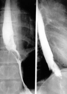
  • Barium swallow showed an intraluminal mass at the esophagogastric junction.

Fig. 1. Pre-Operative Barium Swallow (Select Image for High-quality Version). This demonstrates an irregular intraluminal lesion at and above the gastroesophageal junction.

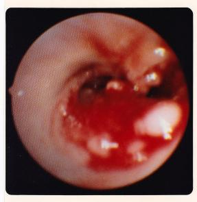
Fig. 2. Endoscopic View of Upper End of Esophagogastric Adenocarcinoma (Select Image for High-quality Version). Esophagoscopy showed an obvious carcinoma of the esophagus, a well differentiated adenocarcinoma histologically.

Treatment Strategy

  • Using a left thoracoabdominal approach, tumor was removed and all lymph nodes in the specimen were negative microscopically for tumor.
  • Patient had an uneventful recovery from the esophagogastrectomy in November 1987.

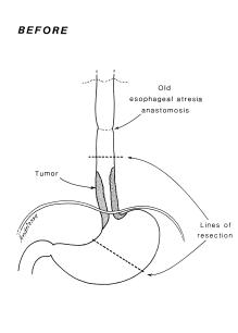
Fig. 3. Pre-Operative Anatomy (Select Image for High-quality Version). Diagram shows the extent of tumor and of resection to be performed using a thoracoabdominal exposure. 

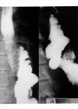
Fig. 4. Post-Operative Barium Swallow (Select Image for High-quality Version). This shows colon interposition from mid esophagus to remaining stomach.

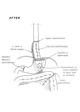
Fig. 5. Post-Operative Anatomy (Select Image for High-quality Version). Anatomy after tumor resection, and restoring gastrointestinal continuity with segment of transverse colon on left gastric pedicle. Note pyloroplasty to enhance gastric emptying.

Follow Up

  • In 1992, 15 years later at age 26 years we performed a left thyroid gland total removal.
  • Histology showed benign modular goiter.
  • Patient last seen in 1994 and she was well.