Browse THP Patient Cases

Urinary Tract Reconstruction Patient 3: Male Infant with Horseshoe Kidney

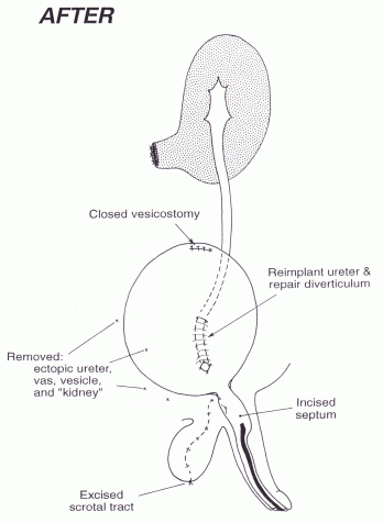
This 1.5-year-old male baby was referred in February 1994 with multiple anatomic problems depicted below in the Before figure. Cutaneous vesicostomy had been performed elsewhere as an infant. Previous studies had shown a horseshoe kidney, the left side of which was dysplastic. The right ureter ended in a retrovesical “cyst.” Because of vesicoureteral reflux, he had been maintained since infancy on prophylactic antibiotics. Prolonged endoscopy, through duplex urethras, and the vesicostomy, plus appropriate radiology studies, disclosed the anatomy seen in the Before figure.

In a 10-hour operation the dysplastic right kidney was removed, together with the retro vesical “cyst” formed by the lower end of the right ureter, and the right seminal vesicle and vas. The right ureter was reimplanted, repairing the adjacent diverticulum as well. The perineal sinus tract connected with the prostatic urethra was excised. The septum between the two urethras was incised endoscopically.

At age 3 years, endoscopic cutting of the septum between his two penile urethras was performed, creating a single urethral meatus, and correcting his mild hypospadias.

Anatomy (Select Image for High-quality Version). (Top) Before undiversion. (Bottom) After undiversion.

Urinary Tract Reconstruction Patient 2: Severe Hydronephrosis in a Solitary Kidney

This 9-year-old girl was referred in February 1972. At age 5 weeks, an evaluation for failure to thrive showed a solitary right kidney with severe hydronephrosis literally filling the abdomen. Pyeloplasty was attempted, but failed. An inlying nephrostomy tube was placed and was being changed monthly.

Bladder volume was only 50 ml. Dilating it hydrostatically, although done gently, ruptured the dome. The dome was closed and pyeloplasty was performed, reducing the size of the renal pelvis. A nephrostogram 10 days later showed an easy passage of contrast to the bladder. Cystogram 4 months later showed increased bladder size, but massive reflux. The ureter was reimplanted curing the reflux. In Dec.1977, because she was small (4 feet, 3 inches tall), she was referred to an Endocrinologist who made the diagnosis of Turner’s Syndrome.

Scoliosis surgery was performed in 1980 at age 18 years. The patient has remained under close metabolic supervision; she has a stable creatinine level of 1.3.

X-rays (Select Image for High-quality Version). (Top Left) Delayed IVP as neonate showing huge right hydronephrosis. (Top Right) Nephrostogram and simultaneous retrograde pyelogram at age 9 years to assess anatomy. (Bottom Left) Nephrostogram 10 days after pyeloplasty. (Bottom Right) IVP age 15 years, note normal bladder size.

Anatomy (Select Image for High-quality Version). (Before) Dismembered pyeloplasty with reduction in size of renal pelvis. (After) Reimplantation of ureter for massive reflux 6 months later.

Urinary Tract Reconstruction Patient 1: Undiversion Case with Cloacal Anatomy

This 5-year-old girl was referred in Sept. 1980 for extensive pelvic surgery. More than half her life since birth she had been hospitalized elsewhere. She had been born with cloacal anatomy with high confluence of her bladder, two vaginas, and rectum. The right kidney had a nephrostomy tube in place because the reimplanted right ureter was obstructed. The left ureter also had an obstructed ureteral reimplant and drained via a loop ureterostomy. The bladder was severely scarred.

The first procedure was to create a urethra and bring the two vaginas to the perineum. The left ureterostomy was closed, its lower end was reimplanted, and a right to left transureteroureterostomy was done. Six months later the bladder, which did not enlarge with hydrostatic stretching, was augmented with ileum.

This case illustrates the trouble which can be created when the surgery gets started “in the wrong direction.” High vaginas require a pull thru procedure, disconnecting them and using the UG sinus as a urethra. Her early years were a nightmare existence and lost most of her renal function.

With a devoted mother to lead her, the child entered adolescence as a young woman with self assurance, doing well socially and psychologically. 

Anatomy (Select Image for High-quality Version). (Before) Preoperative anatomy when referred at age 5 years. Note prior reimplanted ureters, both obstructed. Right kidney drained by nephrostomy tube; left by loop ureterostomy. (After) Initial surgery to fashion a urethra from what had been the urogenital sinus and covered with flap from buttocks. After undiversion 6 months later by left long tunnel reimplant, and transureteroureterostomy; and ileal augmentation to increase capacity of scarred and contracted bladder.

Patient X-rays (Select Image for High-quality Version). (Top Left) Antegrade perfusion of kidneys initially. (Top Right) Showing reconstructed urethra. Malformation: Confluence-Hi (Bottom Left) and (Bottom Right) Augmentation.